
So, what’s noted palaeoartist Brian Engh been up to lately? Well, last Thursday (February 7) JAMA Oncology published Triassic Cancer – Osteosarcoma in a 240-Million-Year-Old Stem Turtle (link) by Haridy, Witzmann, Asbach et al. The paper documents the presence of a malignant tumour present on the femur of a Triassic stem-turtle, Pappochelys rosinae. Although the specimen resides in the collections of the Staatliches Museum für Naturkunde in Stuttgart, it underwent analysis at the Museum für Naturkunde Berlin, where lead author Yara Haridy is based. (In fact, it was “macroscopically investigated”, a CT scan performed, and the 3D data analysed using what must be really cool kit.) The authors’ diagnosis, based on a number of factors detailed in the paper, was “periosteal osteosarcoma”, or bone cancer on the outer surface of the bone. This would be an intriguing find in any Mesozoic animal, but what’s so significant in this case is that it’s the oldest known instance of cancer in an amniote, offering an important insight into the evolutionary history of the disease.
So, Brian’s job was to draw a stem-turtle with a squiffy leg.
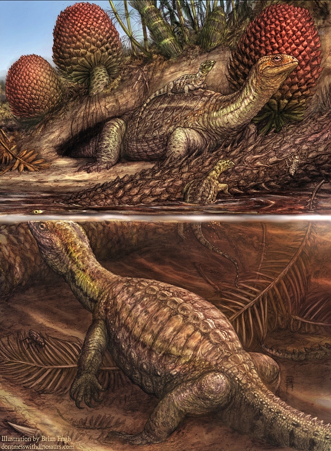
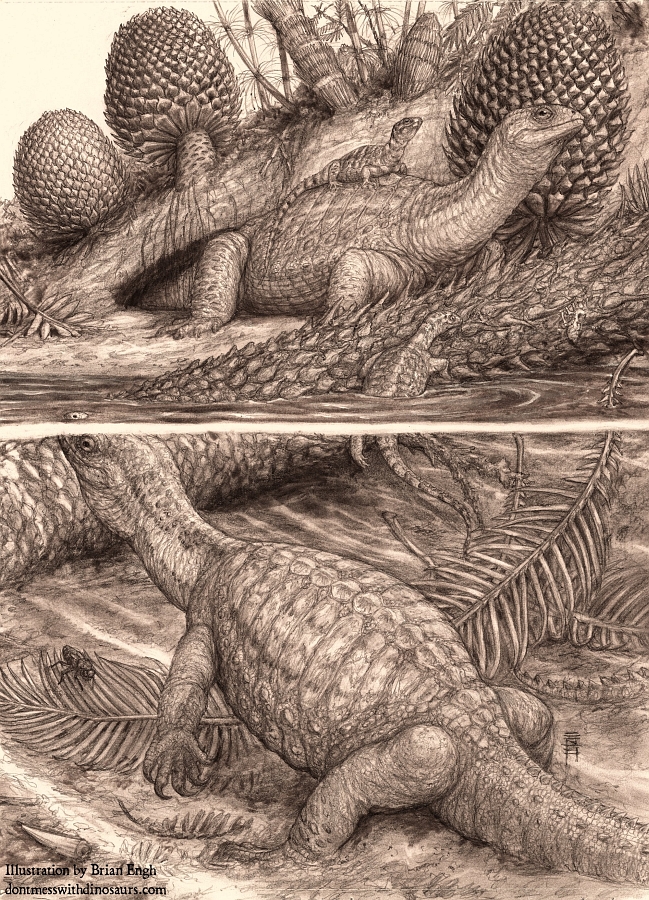
All art is © Brian Engh, used with permission
I’m kidding, of course. What really struck me about Brian’s piece – shown above – was that he envisaged a complete palaeoenvironment where so many others would simply have painted the stem-turtle in question and called it a day. And to be fair, we probably wouldn’t have blamed them. A painting of Pappochelys with a diseased leg – what more do you want? That Brian provides a complete scene with individuals of varying ages and appropriate fauna and flora is truly commendable. But why go to such lengths? I put this to Brian, who in his usual way, shrugged and stated simply, “I try”.
I’m kidding again. In true Brian style, he was highly enthusiastic in answering my query, such that I’ll have to edit his response down a bit here, much as I am loathe to. In short, he had three sci-comm goals with this piece:
- To show what the animal may have looked like in a way that the general public could understand; probably semiaquatic and not quite a turtle.
- To somehow show an animal with internal cancer.
- To communicate how insanely old Pappochelys is (around 240 Ma!), and therefore how old osteosarcomas (bone cancers) are.
As far as the first goal goes, while the specimen in the study is an isolated femur, Pappochelys‘ skeleton is near-completely known thanks to a wealth of specimens from Germany. Therefore, Brian had a very good grounding for his reconstruction, with the important sci-comm decisions coming in his restoration of the soft tissues. Says Brian: “We do know it had expanded ribs with a grooved surface texture that reminded me of the texture that some turtles, such as snapping turtles, have on their skulls in areas with expanded and thickened keratinous scales, so I went with an interpretation showing something like a ‘proto shell’ of expanded keratinous scutes closely hugging the ribs, but not yet thickened enough to have developed into osteoderms with their own bony cores.” Thus, it’s instantly recognisable as a not-quite-turtle. I quickly noted the dopey turtle-like facial expression, too.
The second goal was trickier; prehistoric animals with diseases are rarely portrayed in palaeoart, never mind internal diseases. Here’s where the other Pappochelys individuals come into play; they are healthy and vibrant, while the sick individual is shown to be suffering, albeit subtly. “Yara informed me that in many cases osteosarcomas (bone cancers) metastasize (spread) to the lungs, so our sickly turtle is just hanging in the water trying to breathe.” The youngsters also make it clear that the diseased individual was an adult, and the gathering of stem-turtles reflects bonebeds consisting of differently-aged individuals, which indicate that Pappochelys might have been social.

When it came to the third goal, Brian simply wanted to convey the dizzying strangeness of the Triassic world within this small section of it – and simultaneously link it to the modern day via the unfortunate stem-turtle. Readers of this blog will be familiar with how bizarre the animals were in the Triassic, but Brian wanted to make it clear that the plants, too, would look completely alien to a human. I’m going to have to quote him at length, because he doesn’t half paint a picture, so to speak.
“The environment [of Middle Triassic Germany] was perhaps even stranger than the animals, as many of the dominant plant groups growing there are now totally extinct, with only a few seemingly tame members of related clades alive today. The horsetails grew as large as giant bamboo. The conifers looked like they were made of huge prickly ostrich feathers. Cycads and their extinct sister clan the Bennettitales grew in the familiar semi-arborescent forms as well as in wildly bifurcating feathery leaved bushy forms and even creeping vine-like forms. Perhaps most alien of all, there were a diversity of clubmosses – the evolutionary intermediary between simple mosses and proper vascular plants. Some of these clubmosses grew as large as small trees, and others, such as the Lepacyclotes featured prominently in my scene were apparently adapted to handling the semi-saline conditions in these brackish coastal swamps.”
In showing an animal suffering from a familiar disease in this utterly unfamiliar and incomprehensibly ancient setting, Brian hoped to encourage viewers to consider our evolutionary links to animals like Pappochelys, and also just how long cancer has been afflicting us amniotes.
Brian’s work here is quite important as a rare example of palaeoart depicting an animal with an internal disease, based on a real specimen. There are plenty of other prehistoric animal specimens out there showing examples of pathologies and diseases that would have been largely invisible in life, but it takes a particular skill, thoughtfulness and subtlety to portray them effectively. (There’s definitely a discussion to be had there!) That Brian managed to make this illustration so much more than just a lizard-turtle with a lumpy leg is surely to be applauded.
And while I’m at it – thanks are due to Brian and Yara for contacting us about this discovery and for answering my questions. If you haven’t already, check out the paper and Brian’s own post on his blog, dontmesswithdinosaurs.com.


2 Comments
Andrew Stuck
February 19, 2019 at 11:34 amBrian Engh continues to wow me! That he can communicate so much in a single image is simply stunning.
Thomas Diehl schreibt: Sieben am Sonntag 03.03.2019
March 3, 2019 at 3:08 pm[…] Entdeckung, A einem in Stuttgart gesammelten Skelett der Urschildkröte Pappochelys haben Wissenschaftler einen Knochentumor gefunden. Knochenkrebs ist so ziemlich der einzige Krebs, der sich bei Fossilien leicht nachweisen lässt, […]